
Ultra-high field mechanisms of sensory adaptation across cortical depth
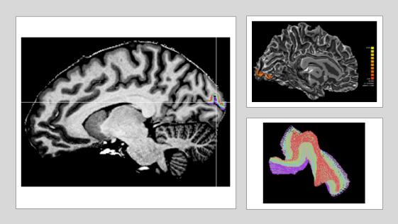 High resolution functional magnetic resonance imaging (UHF-MRI) allows us to investigate cortical depth dependent responses in specific brain areas. Here, we use 7T fMRI to study the laminar organisation of sensory adaptation: a plasticity mechanism involving changes in perceptual sensitivity and brain activity due to continuous sensory stimulation. The high spatial resolution that UHF imaging provides (0.8mm isotropic voxels), allows to investigate the cortical circuits involved in visual adaptation; that is, feedforward mechanisms involved in processing of incoming sensory information vs. feedback mechanisms related to prediction errors (i.e. difference between stimulus expectation and sensory input) from cortical areas higher in the processing stream. Thus, the advantageous spatial resolution of UHF-fMRI, provides a mean to unravelling the brain circuits underpinning our ability to adapt to familiar signals and select novel sensory information.